
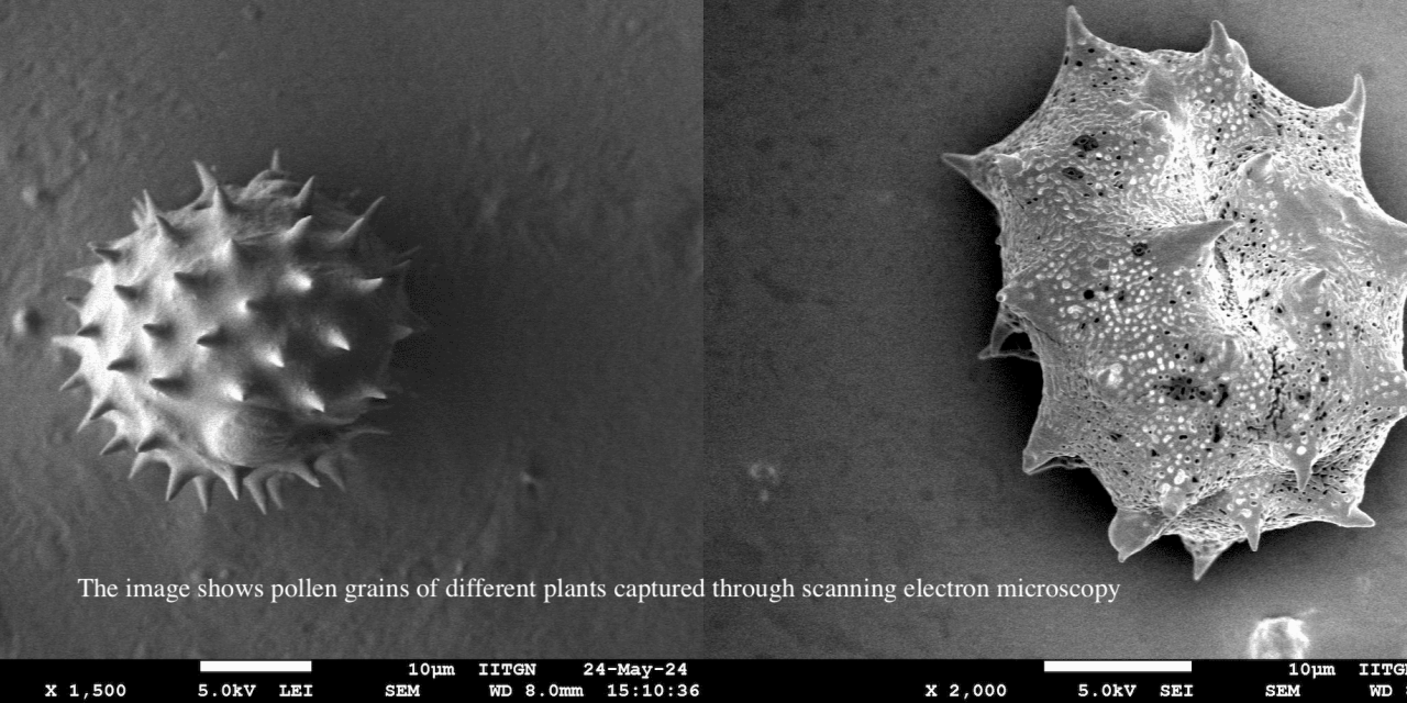
| STORY CREDITS Writer: Apeksha Srivastava Photo: From the research paper “A computer vision methodology for pollen classification using SEM: a case study with medicinal plant species” |
Have you ever seen a hibiscus flower? Although its petals have a range of colours, what makes the trumpet-shaped flower more beautiful is the central stalk, which houses the anthers that produce pollen grains. Powdery in structure, these pollen are commonly bright yellow or golden in colour. During my childhood, I often touched the stalks of these fascinating, bright red flowers, which caused the ‘golden dust’ to stick to my fingers!
Fine dust: This is exactly what pollens may look like at first glance if they are large enough. However, most plant pollens are microscopic, meaning they are invisible to the naked eye. Why is it important? Well, it facilitates reproduction in plants. Further, the morphology of pollens, which refers to their shape, size, and microscopic surface features, can help identify and classify different plant species. Interestingly, this morphology is very diverse, including dotted patterns, boats, cups, and prisms, to name a few. Palynology, which involves the scientific study of pollen grains among other microscopic particles, can use pollen morphology for wide applications ranging from reconstructing historical climate change to examining pollen-related allergies for improving public health responses and monitoring biodiversity. Imagine the depth and diversity of information these teeny tiny pollen grains can provide – interesting and awesome, isn’t it?
Working in this direction and as an effort towards automated and time-efficient analysis of pollens based on their morphology, researchers from the Indian Institute of Technology Gandhinagar (IITGN) presented an approach that combines scanning electron microscopy (SEM) with artificial intelligence (AI). Their findings were recently published in Botany Letters. SEM uses a focused electron beam to visualise microscopic surface structures in detail, which makes it a perfect process to analyse pollen grains. Further, using a representative dataset of SEM images from 28 diverse medicinal plant species, the team developed a computer vision model and a web-based application that isolated pollens from images and identified their associated plant species with remarkable accuracy.
“Although robust imaging techniques are available, standardised workflows and large-scale datasets to facilitate automated pollen analysis are still lacking, especially within underrepresented plants,” explained Jaidev Sanjay Khalane, a final-year undergraduate from IITGN and the first author of the study. Mr Khalane completed this study as part of IITGN’s flagship Summer Research Internship Program (SRIP) with Dr Subramanian Sankaranarayanan, Assistant Professor in the Department of Biological Sciences and Engineering and the Principal Investigator at the Plant Molecular & Developmental Cell Biology (PMDCB) Laboratory.
“The team began by collecting 28 distinct flowering plant species with known medicinal properties from the IITGN campus in Gujarat, India. Pollen analysis using SEM at the Institute’s Central Instrumentation Facility revealed distinct and unique features in pollen shape and surface, ranging from spheroid to irregular and smooth textures to spiny projections,” said Dr Sankaranarayanan. This was followed by the development of MPalyn (Medicinal Pollen and Palynology SEM) database. This web-based, open-access application includes plant species details and corresponding high-resolution SEM images for structured exploration. Such shared resources are critical, and open databases may allow researchers worldwide to constructively build upon the existing information available in their fields. While its current dataset includes medicinal plants with 269 images for segmentation and 5842 images for classification, the framework can also be adapted for future extensions.
As the next step, the team implemented YOLOv11n (You Only Look Once)! It is a computer vision model to extract high-resolution pollen grain images with minimal background noise from SEM images. One can think of it as something that is trained to filter pollen grains, ignoring everything else in an image. Computer vision is a class of artificial intelligence that trains computers to “see” and make sense of that visual information, such as recognising patterns. “This method can be used in large-scale studies and reduces dependency on the time-consuming and difficult-to-scale manual segmentation,” added Dr Nilesh Gawande, former postdoctoral fellow at the PMDCB lab and Assistant Professor at Woxsen University, Hyderabad.
The researchers also tested multiple classification models on manually segmented pollen images. They found that the Vision Transformer (ViT) model achieved the highest classification accuracy. It means that this model effectively learned and discriminated minute differences in pollen structures and correctly identified pollen grains from every species it was shown. According to Dr Shanmuganathan Raman, “Overall, the study involves an interdisciplinary methodology by integrating microscopy and computer vision models to accurately and speedily automate pollen analysis, with broad applications in domains ranging from plant taxonomy to agriculture, medicine, and paleoecology.” Dr Raman is a Professor at the Department of Computer Science & Engineering (CSE) and Electrical Engineering. He is also the Head of the CSE department and the Principal Investigator at the Computer Vision, Imaging, and Graphics (CVIG) Lab.
The researchers noted that improving models and algorithms can help decrease the chances of error in classifying pollens that are morphologically similar or belong to closely related species. This research effectively demonstrates how machine learning can be trained to interpret nature’s tiniest imprints.
The team acknowledged the Department of Biotechnology for the Ramalingaswami re-entry fellowship grant and a start-up grant from IITGN to Dr Subramanian Sankaranarayanan. Discussing the SRIP opportunity, Mr Khalane expressed, “Getting the opportunity to be a first author on a research paper is a great experience, especially when you are an undergraduate student. IITGN’s SRIP exposes students to high-quality and impactful research under the mentorship of experienced faculty. This project has deeply shaped my understanding of my own potential and my future career decisions.” In the words of Dr Sankaranarayanan, “SRIP provides an environment where students can move beyond textbooks. Such research experiences are essential for capacity building as they inculcate critical thinking and scientific confidence in students. Such initiatives contribute towards the research ecosystem by preparing students for interdisciplinary and innovation-driven careers. It is not just about individual training, but also concerns building long-term research capacity.”
*************************
Information on this research has been covered by the following platforms:
- Phys.org
- Press Information Bureau
- Nature India
- The Times of India
- The Live Ahmedabad
- VNI News
- Bio Patrika
- BW Education
It has also been published in print by The Times of India, Divya Bhaskar, and Patrika.